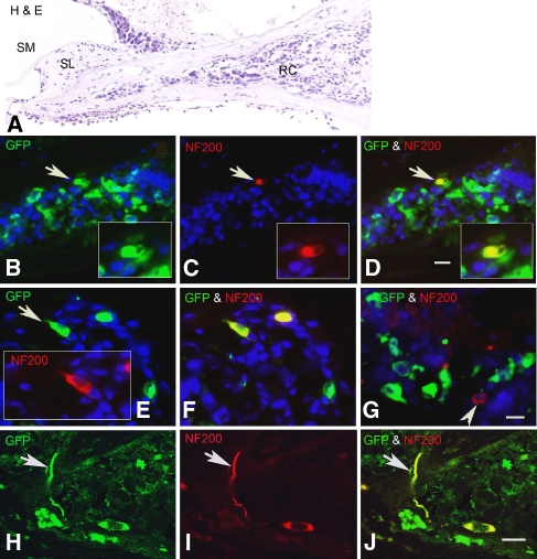
FIG. 7.
Neuronal differentiation of transplanted ESCs in RC of early post-injury cochleas. All sections were obtained from two cochleas 3 weeks after transplantation with GFP-expressing ESCs. Dual immunostaining for GFP (green) and NF 200 (red) antibodies was used to identify ESCs differentiating towards a mature neuronal phenotype. A Radial section stained with H&E shows the profile of the RC transplanted with ESCs. B–D Double staining with GFP and NF 200 antibodies shows an ESC (arrow) that has differentiated into a NF-200-positive neuron-like cell (yellow). The section is about 20 μm apical to the one shown in A. E, F Section from another transplanted ear showing two NF-200-positive cells differentiated from ESCs. G A native neuron (arrowhead) remaining in RC is not GFP-positive. H–J Confocal images of a radial section stained for GFP and NF 200 antibodies show a neuron-like cell with its process (arrow) differentiated from an ESC. The section was taken from the ear shown in sections E and F. SM scala media, SL spiral limbus. Scale bar = 15 μm.

