FIG. 3.
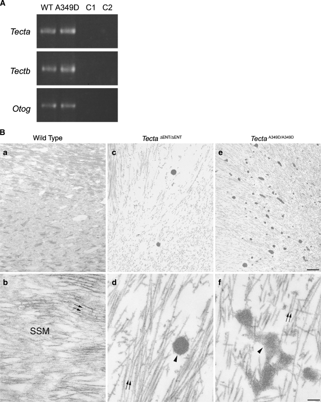
Reverse transcriptase PCR analysis and ultrastructure of the tectorial membrane. A Tecta, Tectb, and Otog messenger RNAs (mRNA) are expressed in TectaA349D/A349D cochleae. Randomly primed first-strand cDNA was prepared from total cochlear RNA from Tecta+/+ (WT) and TectaA349D/A349D (A349D) mice, and products specific for Tecta, Tectb, and Otog mRNAs were amplified by PCR. C1 Reverse transcriptase reaction control, in which the reverse transcription reaction was performed without reverse transcriptase and then amplified by the PCR, C2 control PCR performed without an aliquot of the reverse transcriptase reaction. B Electron micrographs illustrating the ultrastructural appearance of the tectorial membrane in wild-type (a, b),  (c, d), and TectaA349D/A349D (e, f) mice at postnatal days P80, P77, and P82, respectively. Collagen fibrils (arrows) are embedded in a striated-sheet matrix (SSM) in the tectorial membrane of wild-type mice (a, b). In TectaΔENT/ΔENT (c, d) and TectaA349D/A349D (e, f) mice, the striated-sheet matrix is absent, and large electron dense granules (arrowheads) are visible. These granules are more numerous in the TectaA349D/A349D mouse than in the
  mouse (compare c and e). Scale bars for a, c, e = 1 μm, for b, d, f = 200 nm.

