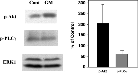
Fig. 6.
Western blot analysis of Akt(PKB) and PLCγ phosphorylation in organ of Corti explants. Explants were treated with 50 μM gentamicin (GM) for 6 h. Phospho-Akt and phospho-PLCγ were detected by Western blot. Total Erk1 was used as an internal control. Phospho-protein levels were determined by densitometry and were normalized against Erk1. Gentamicin treated levels are expressed as % of control values. Phospho Akt was significantly increased by gentamicin treatment (p < 0.05), whereas phospho-PLCγ levels were not.

