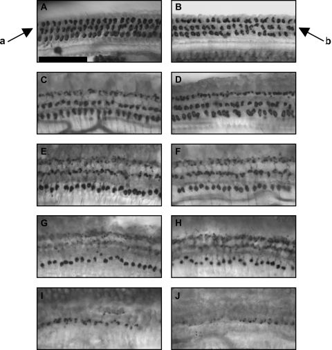
Fig. 2.
Medial olivocochlear (MOC) immunolabeling was not affected by MPTP. Tissues were immunolabeled with antisynaptophysin, and are from base (A, B), second (C, D) and third (E, F) turns, lower apex (G, H) and upper apex (I, J). Control tissues are depicted in the left panels (A, C, E, G, H). MPTP-treated tissues are depicted in the right panels (B, D, F, H, J). Arrows indicate normal MOC immunolabeling below the outer hair cells (arrow a), and unchanged immunolabeling of MOC puncta after MPTP treatment (arrow b). Scale bar = 50 μm.

