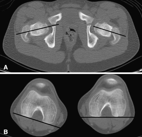
Fig. 10A–B.
A computed tomography scan shows the femur for femoral anteversion with cuts through the hips (A) and distal femora (B) with angle formed by lines drawn through the axis of the femoral neck and femoral condyles. There was internal rotation of the right knee but satisfactory anteversion bilaterally.

