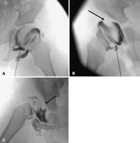
Fig. 5A–C.
(A) A normal hip arthrogram demonstrates thin rim of contrast surrounding the femoral head and labrum covering the femoral head laterally. (B) A subluxated hip with acetabular dysplasia, pooling of contrast medially, and deflected labrum (arrow) is shown. (C) A dislocated hip with inverted labrum (arrow), narrowing of the joint capsule, and acetabular dysplasia is shown.

