Fig. 1.


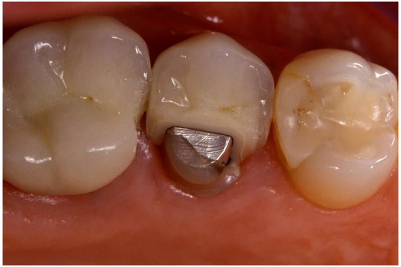
Fig. 1a. Procera AllCeram upper right second premolar crown fracture exposing part of tooth structure and amalgam restoration (Photo: courtesy of Dr. U. Brodbeck, Zurich, Switzerland).
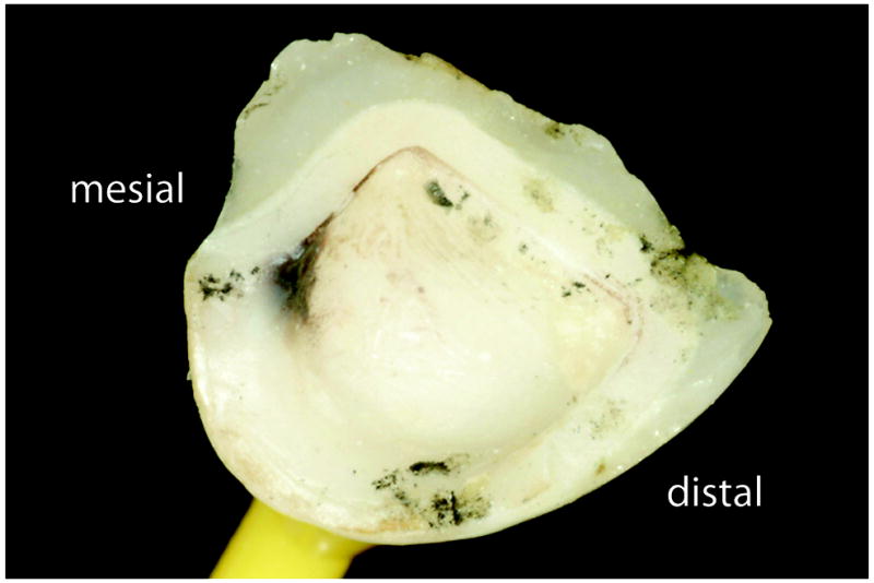
Fig. 1b. Recovered part by the patient of the broken Procera AllCeram premolar crown.
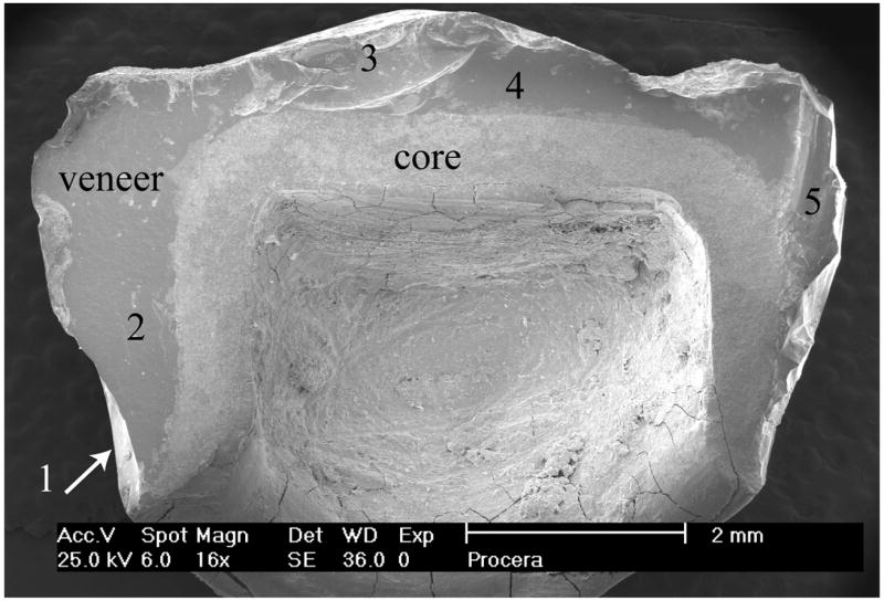
Fig. 1c. Recovered fractured Procera AllCeram crown part viewed at 16× under the SEM. Zones of interest for detailed fractographic analysis are numbered 1 to 5 starting at the mesial (left) margin to the other side of the crown.
