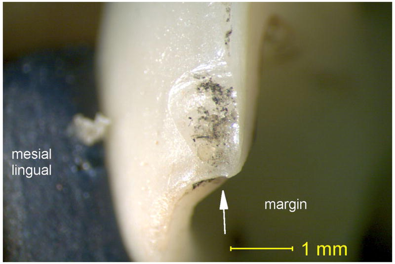
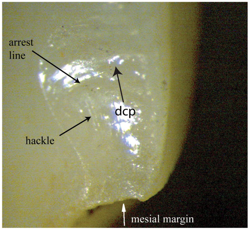
Fig. 2.


Fig. 2a. Stereomicroscope image of a mesial marginal edge chip within the veneering ceramic in zone 1 before cleaning the specimen. Note the localized staining covering the chipped ceramic surface.
Fig. 2b. Stereomicroscope image of the same chipped zone 1 after cleaning the crown part 10 minutes in an ultrasonic ethanol bath removing the stain. The arrow labeled “dcp” shows the direction of crack propagation.
