Fig. 3.


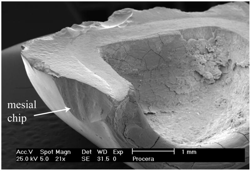
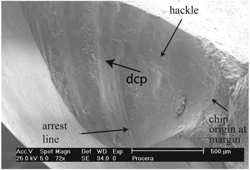
Figs. 3a-c. SEM images of the chip in zone 1 at different magnifications. Arrest lines, hackle and wake hackle are recognizable and provide the direction of crack propagation (dcp) as marked by a black arrow from the margin upwards.


Figs. 3a-c. SEM images of the chip in zone 1 at different magnifications. Arrest lines, hackle and wake hackle are recognizable and provide the direction of crack propagation (dcp) as marked by a black arrow from the margin upwards.