Fig. 4.


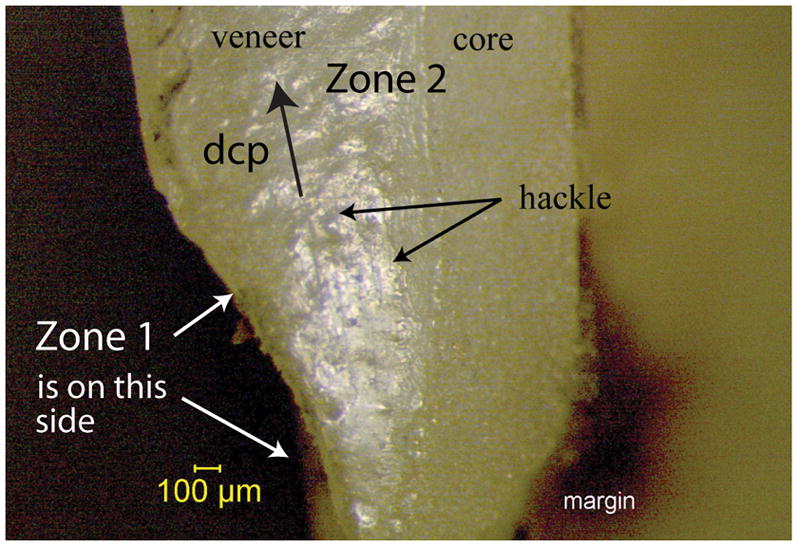
Fig. 4a. Stereomicroscope image of zone 2 of interest. Parallel running hackle lines are concentrated in the veneering ceramic. These indicate the direction of crack propagation is from the bottom to top in this view.
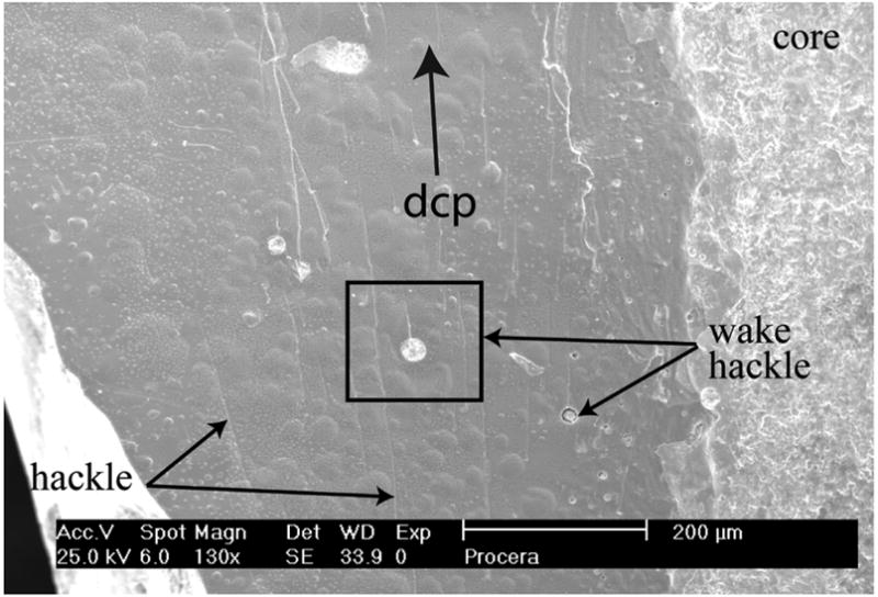
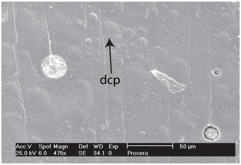
Figs. 4b,c. Same area as in Fig. 4a viewed under the SEM at higher magnification. Hackle and wake hackle (emanating from pores and inclusions) are easily distinguished and used to indicating the crack was running upwards towards the occlusal side.
