
Figure 6. 3-O-sulfated octasaccharide inhibits HSV-1 entry.
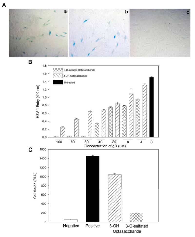
Panel A shows the nhibition of HSV-1 entry into cultured corneal fibroblasts (CFs). CFs were infected with 1PFU (plaque-forming units)/well of HSV-1(KOS-gL86) preincubated with buffer alone (a), 100 μM of 3-OH octasaccharide (b) or 100 μM of 3-O-sulfated octasaccharide (c). At 6 hrs later, the cells were washed, fixed and incubated with X-gal to identify infected cells (dark cells). Panel B shows the dose dependent inhibition of HSV-1 entry into HeLa cells. HeLa cells were infected with 1PFU (plaque-forming units)/well of HSV-1(KOS-gL86) preincubated with indicated concentration of 3-OH octasaccharide or 3-O-sulfated octasaccharide as shown. Untreated cells were used as a control. About 6 hrs post infection the cells were lysed for the quantification of β-galactosidase activity as a measure of viral entry. Absorbance at 410 nm (OD 410 nm) of ONPG reaction products were plotted against the concentration of the octasaccharides used. Panel C shows the specific inhibition of HSV-1 glycoproteins-induced membrane fusion. CHO-K1 cells were used as effector and target cells. Effector cells were transfected with plasmid expressing HSV-1 glycoproteins and luciferase reporter plasmids. Target cells were transfected with T7 RNA polymerase and the plasmid expressing 3-OST-3. Luciferase activity was measured 24 hrs after mixing and co-cultivating the effector and target cells. The luciferase activity is from one experiment performed in triplicate. The concentrations of 3-OH or 3-O-sulfated octasaccharides used in this experiment were 100 μM.