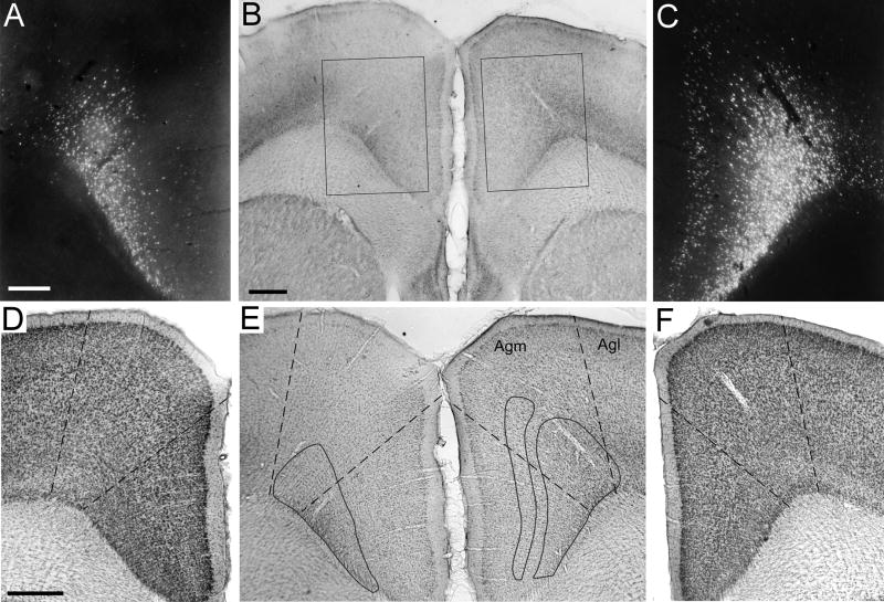
Fig. 13.
Labeled neurons in MI cortex produced by a deposit of FG in the right thalamus. A: Retrogradely-labeled neurons in deep layer VI of the contralateral MI cortex. Scale, 250 μm. B: Unstained section through the left and right MI cortices; rectangles indicate the regions shown in panels A and C. Scale, 500 μm. C: Retrogradely labeled neurons in layers Vb and VI of the ipsilateral MI cortex. Same magnification as panel A. D: Thionin-stained section through left MI cortex located adjacent to the section in panel A. Dashed lines indicate the borders of the medial agranular (Agm) cortex. Scale, 500 μm. E: Same section as in panel B but shown at magnification used for panels D and F. Solid contours show locations of retrogradely-labeled neurons after superimposing the fluorescent photomicrographs in A and C onto the section. Dashed lines show approximate Agm borders after superimposing the thionin-stained sections in panels D and F onto the section. F: Thionin-stained section through right MI cortex located adjacent to the section in panel C. Dashed lines and magnification are shown as in panel D.

