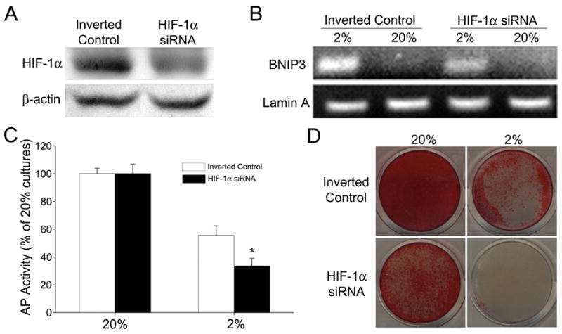
Figure 6.

Effect of HIF-1α silencing on AP activity and mineralization of MLO-A5 cells. (A) Western blot analysis of HIF-1α siRNA transfected MLO-A5 cells and cells transfected with inverted controls after 6 h at 2% O2. HIF-1α protein expression was reduced in the silenced cells. (B) RT-PCR analysis of the HIF-1α target gene, Bnip3. There was reduced expression of Bnip3 in the silenced MLO cells maintained at 2% O2 for 8 h. (C) AP activity of MLO-A5 cells after 7 days of culture at 20 or 2% O2. There was a significant reduction in activity in the HIF-1α silenced cells at 2% O2. Activity is expressed as ng/min/μg protein and normalized to 20% O2. Values plotted are mean and SEM. * denotes significantly different from cells transfected with the inverted control at 2% pO2; p<0.05. (D) Alizarin Red staining of silenced MLO-A5 cells after 7 days in culture at 20 or 2% O2. HIF-1α silenced cells displayed a reduction in staining at both O2 tensions.
