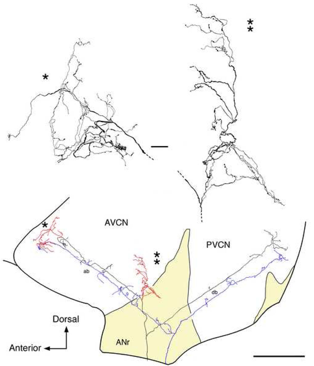
Figure 1.
Drawing tube reconstructions of a low SR auditory nerve fiber (black and red, CF=3.1 kHz; SR=0.2 s/s; Th=26 dB SPL) and a high SR auditory nerve fiber (blue, CF=1.2 kHz, SR=86 s/s; Th= −3 dB) as viewed laterally. These fibers are from separate cats but are shown together to illustrate the differences in branching patterns. Their placement in the nucleus is based on tonotopic maps of the fibers (Ryugo and May, 1993; Ryugo and Parks, 2003) and cochlear nucleus (Bourk et al., 1981). The ascending branches take a relatively straight trajectory through the AVCN. Low SR fibers are distinctive by the collaterals that arborize within the small cell cap (red). Otherwise, the main parts of the ascending and descending branches are similar for the different SR fiber types (black, low SR; blue, high SR). Higher magnification drawings are shown for each collateral. One collateral ramifies anterior to the endbulb (*), whereas the other ramifies laterally (**). The ascending branch has been modified from Fig. 15 of Fekete et al., 1984 in order to show details of the collateral branching. Abbreviations: ab, ascending branch; ANr, auditory nerve root; AVCN, anteroventral cochlear nucleus; db, descending branch; PVCN, posteroventral cochlear nucleus. The scale bar equals 25 µm for the high magnification collateral drawings (top) and 0.5 mm for the orientation drawing (bottom).

