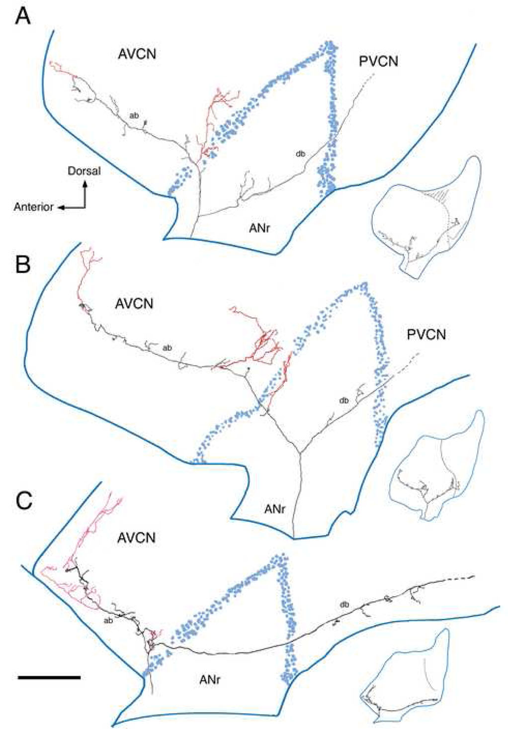
Figure 2.
This figure presents three reconstructed low SR fibers in the cochlear nucleus in side view. The collaterals that innervate the small cell cap rostrally and laterally are shown in red. (A) CF=1.2 kHz, SR=1.0 s/s, Th=4 dB SPL; (B) CF=1.85 kHz, SR=0 s/s, Th=50 dB SPL; (C) CF=0.3 kHz, SR=0.1 s/s, Th=39 dB SPL. The distributed terminals from these collaterals have areal spread over the nucleus yet are also confined to a thin zone squeezed between the outermost GCD and the underlying magnocellular core. Abbreviations: ab, ascending branch; ANr, auditory nerve root; AVCN, anteroventral cochlear nucleus; db, descending branch; PVCN, posteroventral cochlear nucleus. Scale bar equals 0.5 mm.

