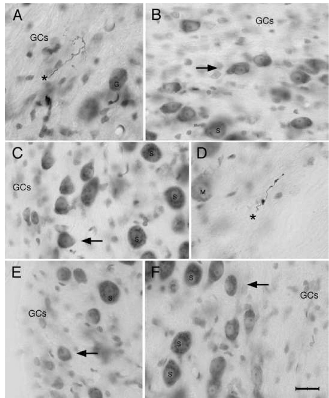
Figure 4.
Photomicrographs of HRP-labeled collaterals from low SR auditory nerve fibers (A, D asterisks) and small cells of the cap stained with cresyl violet. Note that the labeled fibers are thin and the swellings are small (A, CF=0.17 kHz, SR=0.06 s/s, Th=26 dB SPL; D, CF=2.8 kHz, SR=1.6 s/s, Th=29 dB SPL). The fiber in A was montaged and pieced together across multiple focal planes. The small cells (arrows, B, C, E, F) have a qualitatively similar appearance in addition to their small size. They have a pale, round, and slightly eccentric nucleus with a prominent nuclear envelope. Even though they vary somewhat in size and shape, the cytoplasm and nucleus have a homogenous appearance across the population. These cells were selected from different locations around the cochlear nucleus. Clockwise: (A) dorsolateral, (B) dorsal, (D) dorsomedial, (F) medial; (E) ventrolateral, and (C) middle lateral. Abbreviations: GCs, granule cells; G, globular bushy cell; M, multipolar cell; S, spherical bushy cell. Scale bar equals 20 µm.

