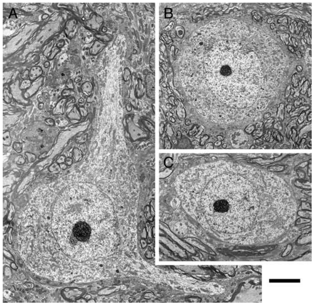
Figure 5.
Electron micrographs of representative small cells of the peripheral cap. Cell bodies appear polygonal when dendrites emerge (A) but are oval-to-round when no dendrites are evident (B, C). The nucleus is pale compared to the cytoplasm. Somatic terminals are generally sparse but increase around the base of dendritic stalks. Scale bar equals 5 µm.

