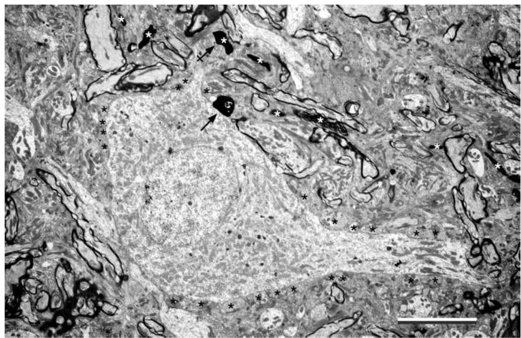
Figure 6.
Electron micrograph of a small “polygonal” cell in the dorsal small cell cap that is innervated by a low SR fiber (CF=0.78 kHz; SR=6.33 s/s; Th=20 dB). The cytoplasmic features resemble those of the small cell shown in Figure 5A. The labeled terminals arise from a single collateral and make asymmetric synapses on the cell body (arrow) and one of its dendrites (arrow with cross). The PSDs were observed in deeper sections and the vesicles were revealed by “overexposing” the terminal. Unlabeled axosomatic terminals are labeled with black asterisks. Additional labeled terminals and their fiber of origin are indicated (white asterisks). The dendrites are marked by polyribosomes. Scale bar equals 5 µm.

