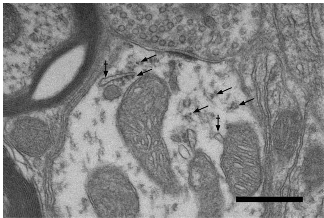
Figure 8.
Electron micrograph of axodendritic synapse in the small cell cap. The terminal contains large, round synaptic vesicles and forms an asymmetric synapse onto a dendrite that contains microtubules, polysomes (arrows), and membranous cisternae (arrows with bar). The presynaptic characteristics are typical of auditory nerve fibers and the postsynaptic dendrite exhibits features typical of small cells of the cap. The positioning of protein synthetic organelles (arrows) beneath dendritic synapses suggests that local translation might play a role in synaptic modification. Scale bar equals 0.5 µm.

