Figure 3.
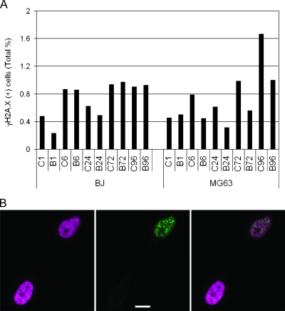
BrdU does not lead to increased γH2A.X immunoreactivity. (A) BrdU-treated (B) and matched control (C) BJ or MG63 cells immunostained for γH2A.X at 1, 6, 24, 72, and 96 hours after a single 24-hour pulse of BrdU, and positive cells are expressed as the percentage of the total cell population. Although there is a trend toward greater γH2A.X expression in both control and treated cells with increasing time in culture, there are no consistent differences between matched control and treated samples in either cell type. (B) Representative photomicrographs showing examples of γH2A.X(+) and γH2A.X(-) MG63 cells 96 hours after a single 24-hour pulse of 50 µM BrdU. The panel on the left shows DAPI staining of two nuclei (pseudocolored magenta). The middle panel shows γH2AX immunolabeling (green) of the same field of view. The right-hand panel is an overlay of two panels. Characteristic γH2AX(+) foci are present in the upper nucleus, indicating double-strand DNA breaks. Scale bar, 10 µm.

