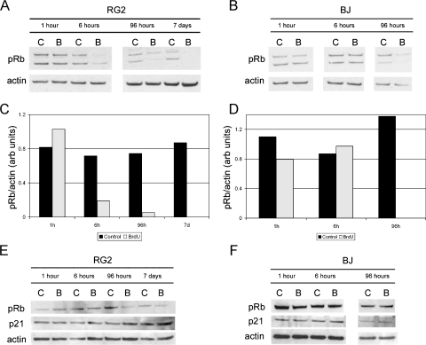
Figure 6.
Phosphorylation of pRb is reduced in some cell types after BrdU exposure, whereas total pRb and p21 remain unchanged. (A and B) RG2 and BJ cells were exposed to a single 24-hour pulse of 50 µM BrdU and analyzed for phosphorylated pRb (Ser249, Thr252) by Western blot analysis at 1, 6, and 96 hours and 7 days after administration. (C and D) Densitometric analysis was performed to quantitate the changes in phosphorylated pRb protein levels in RG2 and BJ samples normalized to actin. There is a dramatic reduction in phospho-Rb beginning at 6 hours in the RG2 cells, and at 96 hours in the BJ cells. (E and F) Similarly treated RG2 and BJ cells were analyzed for total Rb, and p21 protein expression by Western blot analysis. Total Rb decreases slightly in RG2 cells at 1 week after administration but is not altered in BJ cells. In neither cell type is p21 expression altered because of BrdU exposure. (Amount of protein loaded/well: (A and B) RG2 1 hour, 6 hours: 17 µg; RG2 96 hours, 7 days: 20 µg; BJ 1 hour, 6 hours: 15 µg; BJ 96 hours: 17 µg; (C and D) RG2: 15 µg; BJ: 17 µg.)

