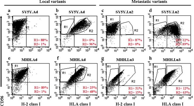
Figure 3.
Immunophenotyping of local and metastatic NB variants. Flow cytometric analyses of the NB local variants, SY5Y.Ad (a, b) and MHH.Ad (e, f), and the NB metastatic variants, SY5Y.Lu2 (c, d) and MHH.Lu3 (g, h), using monoclonal antibodies against CD56/HLA/H-2. Cells were positive for HLA and CD56 but negative for H-2. Percentages of CD56+/HLA+/H-2- were calculated by using the following gating strategy. On a FL1-H/FL2-H dot plot, the CD56+/H-2- cells were identified by region R1. In a second dot plot, region R1 was defined CD56+/H-2-, and region R2 was used to identify the CD56+/HLA+ human NB cells.

