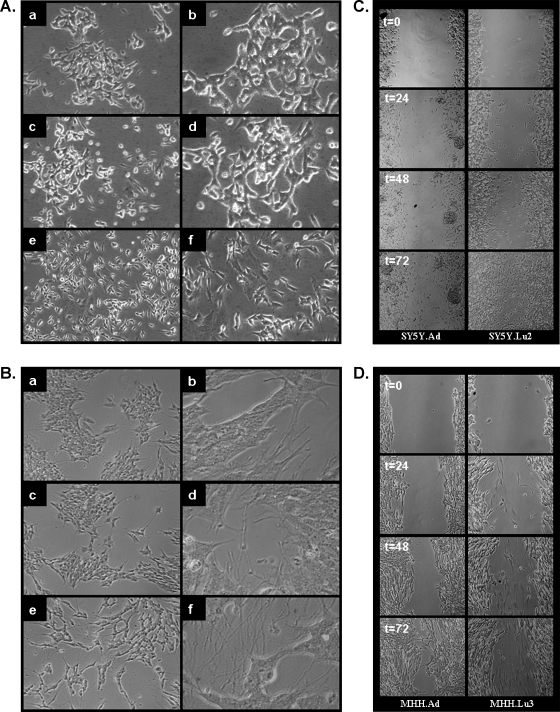
Figure 4.
Distinct cell morphologies and migratory capacity of local and metastatic NB variants in vitro. Morphology of (A) SH-SY5Y (a, b), SY5Y.Ad (c, d), and SY5Y.Lu2 (e, f). Morphology of (B) MHH-NB-11 (a, b), MHH.Ad (c, d), and MHH.Lu3 (e, f). Phase contrast photomicrographs: a, c, and e original magnification, x10; b, d, and f original magnification, x40. Phase contrast photomicrographs of scratched monolayers in a wound healing assay: SY5Y.Ad and SY5Y.Lu2 variant cells (C) and MHH.Ad and MHH.Lu3 variant cells (D) at 0, 24, 48, and 72 hours. Shown are representative photomicrographs (original magnification, x10) of two to three independent experiments.

