Fig. 2.
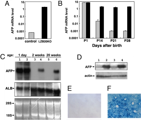
Dysregulated AFP expression in LZB20KO liver. (A) AFP mRNA expression in liver from 4-month-old control (gray bar) and LZB20KO (black bar) mice was shown by real-time RT-PCR. n = 3 experiments. (B) Real-time RT-PCR analysis for AFP mRNA expression in liver from control (gray bar) and LZB20KO (black bar) mice during postnatal days 1–28. Each group at individual time points included at least 5–8 mice. (C) Northern blot analysis for AFP and ALB mRNA expression in the livers from 1-day-, 2-week-, and 20-week-old LZB20KO mice (lanes 2, 4, and 6) and their control littermates (lanes 1, 3, and 5). The RNA loading control was demonstrated by EtBr staining of the gel. (D) Western blot analysis of AFP expression in whole liver lysates from 3-week-old control (lanes 1 and 3) and LZB20KO (lanes 2 and 4) mice. The loading control was demonstrated by reprobing with anti-actin antibody. (E and F) In situ β-galactosidase staining showed LacZ expression throughout the liver from 3-week-old ZBTB20flox/flox/Alb-cre/AFP−/− compound knockout mice (F) rather than the same-aged ZBTB20flox/flox/AFP−/− knockouts (E), which reflected the AFP gene transcriptional activity. Error bar represents SD.

