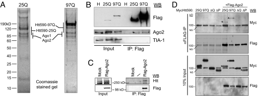
Fig. 1.
Isolation of Flag-tagged WT (25Q) and polyQ-expanded (97Q) Htt protein complexes from HeLa cell cytoplasmic S100 fraction. (A) Eluates from immunopurified Flag-Htt590 resolved by 10% SDS/PAGE and visualized by Coomassie blue staining. Mass spectrometric protein identifications are listed alongside the gel regions where the proteins were identified. (B) Verification of Ago2 as an Htt590 interactor. Immunoblot analysis of Flag immunoprecipitates from the indicated cell lysates probed with the indicated antibodies. (C) Cytoplasmic extract from HEK293T cells stably expressing Flag-Ago2 or Flag-vector (Mock) were immunoprecipitated with α-Flag M2 antibody and probed with α-Htt (HDB4E10, Abcam) or α-Flag antibody. (D) HeLa cells were cotransfected with Myc-Htt590–25Q, -97Q or deletion mutant (ΔQ or ΔP) and Flag-Ago2. Ago2 was immunoprecipitated with α-Flag antibody and the presence of Htt was determined by immunoblotting with α-Myc antibody. *, WT and mutant Myc-Htt590. NS: a nonspecific background band migrating at 100 kDa. Fig. S2D shows clearer separation of Myc-Htt590-ΔQ from the background band.

