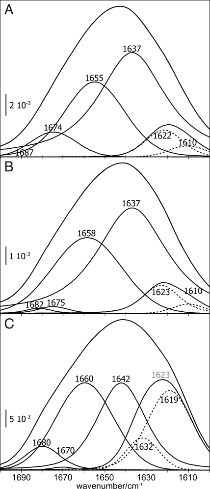
Fig. 2.
The amide I bands of PrPC shift upon membrane anchoring. (A and B) The spectral deconvolution of the amide I band at 0.4 μM PrPC after 34 min incubation with the lipid membrane (A) resembles the one of 1.0 μM PrPC after 3 min (B). (C) After 34 min incubation time at 1.0 μM PrPC, substantial band shifts toward oligomeric β-structure were evident. Bands between 1690–1678 cm−1 were assigned to antiparallel β-sheets, 1,676–1,663 cm−1 to β-turns, 1,662–1,645 cm−1 to α-helices, 1,644–1,635 cm−1 to random coil, and 1,634–1,610 cm−1 to β-sheets. This region was subdivided (dashed bands) approximately at 1,620 cm−1, with the low-frequency component representing intermolecular β-structure. The labels depict deconvolution results, regardless of component intensity.

