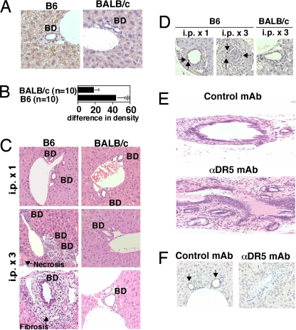
Fig. 2.
Cholangitis induced by anti-DR5 mAb treatment in B6 mice but not BALB/c mice. (A) Immunohistochemical examination of DR5 expression in livers of B6 and BALB/c WT mice. BD, bile duct. (B) Quantification of DR5 expression in immunohistochemistry. Sections were analyzed by using optimal densitometric mean value (MEAND), and data are represented as the mean ± SD (increased % intensity) of plural sections from indicated numbers of mice in each group. ∗, P < 0.05 compared with BALB/c mice. (C) Histological examination of livers in anti-DR5 mAb-treated B6 and BALB/c mice 4 days after one or three injections of anti-DR5 mAb. BD, bile duct. (D) Apoptosis of biliary epithelial cells demonstrated by TUNEL staining. The arrows indicate apoptotic cholangiocytes. (E) Histological examination of extrahepatic bile duct in anti-DR5 mAb- and control Ig-treated B6 mice 4 days after three injections. (F) Cholangiocytes indicated by immunohistochemical staining for cytokeratin 19. The arrows indicate intact bile ducts. Original magnification: ×20 on E and ×40 on others.

