
Figure 4.
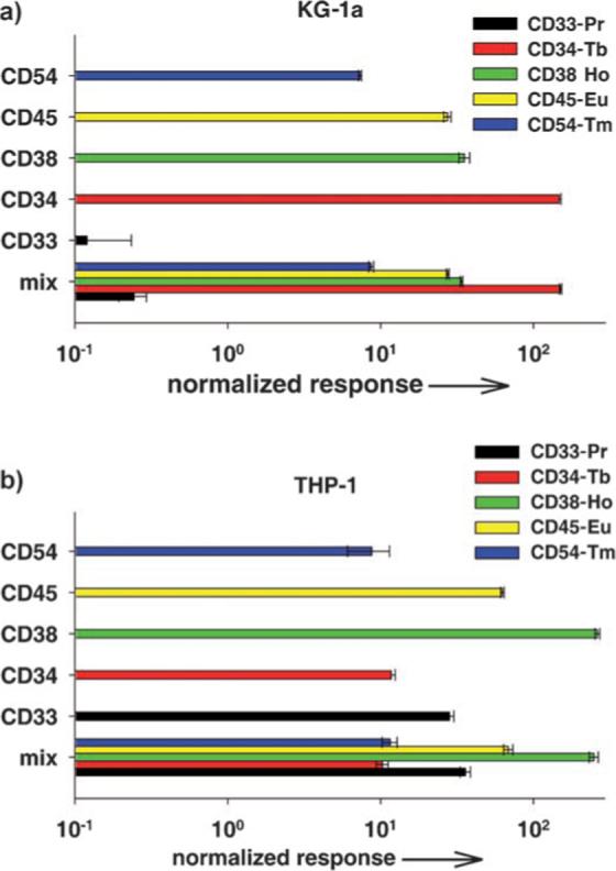
Multiplex analysis of antigen expression by two acute leukemia cell lines. a) KG-1a cells were probed with five element-tagged antibodies to cell surface antigens: CD33-Pr, CD34-Tb, CD38-Ho, CD45-Eu, and CD54-Tm. Background controls included element-tagged mouse IgG-Pr, IgG-Tb, IgG-Ho, IgG-Eu, and IgG-Tm. Triplicate samples with 1 × 10⁶ cells per tube were set up for reaction with each antibody separately and with a mix of all five antibodies together as well as controls. The cells were stained, fixed, and then treated with a RhIII-containing metallointercalator for cell enumeration and signal normalization. Washed cell pellets were dissolved in concentrated HCl, combined with an equal volume of Ir standard solution (1 ppb), and analyzed by ICP-MS. Results are presented as normalized response with respect to Ir, Rh, and background signals from nonspecific IgG binding. b) THP-1 cell line treated as described for KG-1a.