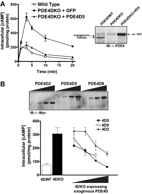
FIGURE 7.
Expression of exogenous PDE4D rescues the phenotype of PDE4DKO MEFs. A, PDE4DKO MEFs were infected with adenoviruses encoding a Myc-tagged PDE4D5 or the green fluorescent protein as a control at a multiplicity of infection of 20 for 36 h. Cells were then stimulated with 10 μm ISO for the indicated times, and cAMP concentrations were measured by RIA. Data represent the means ± S.E. of three experiments and are compared with the cAMP accumulation of uninfected wild type cells. The inset reports a Western blot of extracts prepared from PDE4DWT MEFs, PDE4DKO MEFs, and PDE4DKO cells infected with the PDE4D5 adenovirus using an anti-PDE4 antibody. Similar amounts of protein extract were loaded in all lanes. B, wild type, PDE4DKO, and PDE4DKO MEFs infected with adenoviruses encoding PDE4D5, PDE4D9, or PDE4D2 were stimulated with 10 μm ISO for 5 min. Incubations were then terminated, and cAMP concentrations were measured by RIA. Cyclic AMP accumulation in the presence of exogenous PDE4Ds is compared at different levels of overexpression; at each point the three PDE4D splice forms are expressed at similar amounts as determined by Western blotting (see inset). Data represent the means ± S.E. of three experiments (4D2 and 4D5) or the average and range of two experiments (4D9).

