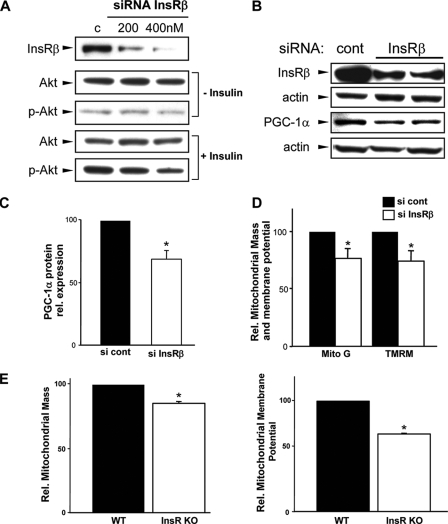
FIGURE 5.
Disruption of insulin signaling and mitochondrial respiration. A, effect of knockdown of InsRβ on Akt phosphorylation (p-Akt). B, PGC-1α levels in response to knockdown of InsRβ. C, quantitative reduction of PGC-1α protein after InsRβ knockdown. D, attenuation in mitochondrial size and membrane potential in insulin receptor knockdown cells. TMRM, tetramethylrhodamine methyl ester; Mito G, MitoTracker green. E, in insulin receptor knock-out (KO) skeletal myoblasts the mitochondrial membrane potential and mitochondrial mass is diminished compared with wild type (WT) control cells.

