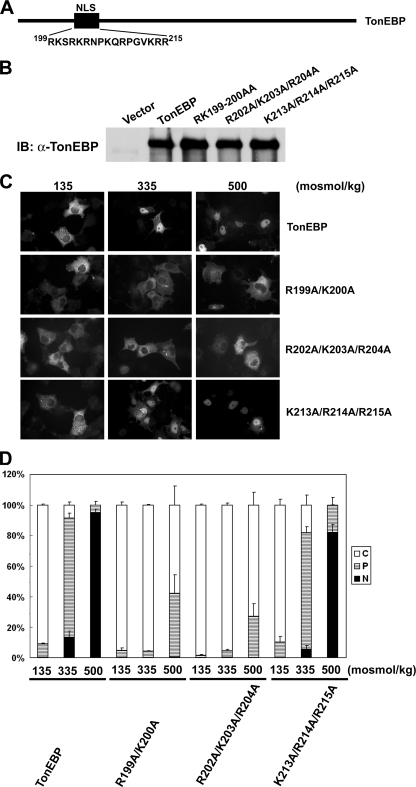
FIGURE 3.
NLS of TonEBP is monopartite. A, schematics of TonEBP and amino acid sequence of residues 199–215. B, immunoblot (IB) of COS7 cells transfected with TonEBP and its site-directed mutants (R199A/K200A, R202A/K203A/R204A, and K213A/R214A/R215A) in fusion with GFP. C, green fluorescence images of the transfected cells that had been incubated for 60 min in hypotonic (135 mosmol/kg H2O), isotonic (335 mosmol/kg H2O), or hypertonic medium (500 mosmol/kg H2O) before they were fixed for observation. D, the cells shown in C were scored as follows: C (open bars) for cells displaying predominantly cytoplasmic distribution of the fluorescence, P (hatched bars) for cells with both cytoplasmic and nuclear distribution, and N (filled bars) for cells with predominantly nuclear distribution. More than 200 cells were analyzed for each condition from a transfection. Values are mean ± S.D. from four independent experiments (n = 4).

