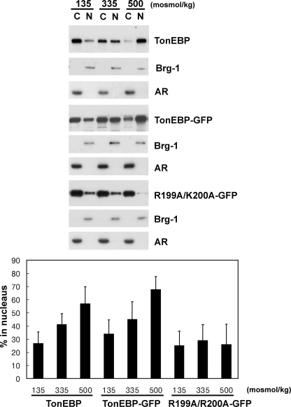
FIGURE 4.
Nuclear distribution of TonEBP, and GFP-fused TonEBP and K199A/R200A. COS7 cells were transfected without DNA (mock) or with expression vector for TonEBP-GFP or R199A/R200A-GFP and then incubated for 60 min in hypotonic, isotonic, and hypertonic medium as described in the legend to Fig. 3. Cytoplasmic (C) and nuclear fraction (N) were obtained from each condition and loaded at a 1:1 ratio for immunoblot analysis of TonEBP (mock-transfected samples) and GFP. In each condition, Brg-1 and aldose reductase (AR) were also immunoblotted to monitor separation of the nuclear versus cytoplasmic fractions. Intensity of TonEBP or GFP signal was quantified and expressed as percentage in the nucleus: 100 × (intensity in N)/(intensity in C + intensity in N) (mean ± S.D., n = 5 or 6). In TonEBP or TonEBP-GFP, all three values are different from each other (p < 0.01, Student's t test), whereas there are no statistical differences in R199A/K200A-GFP.

