FIGURE 5.
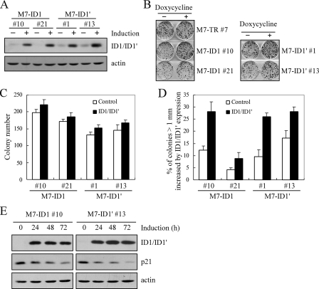
Overexpression of ID1 or ID1′ promotes cell proliferation potentially through inhibition of p21 expression. A, generation of MCF7 cell lines that inducibly express ID1 or ID1′. The levels of ID1 and ID1′ were quantified with anti-ID1. B, ID1 and ID1′ promote colony formation in MCF7 cells. Colony formation assay was performed with MCF7 cells uninduced or induced to express ID1 or ID1′ for 14 days, as described under “Experimental Procedures.” C, quantification of the total number of colonies shown in B. The number of colonies was calculated in triplicate for each cell line. D, quantification of the percentage of colonies with a diameter of >1 mm. The percentage of colonies with a diameter of >1 mm was calculated in triplicate for each cell line. The average was plotted as the percentage of colonies (>1 mm) increased by ID1 or ID1′ expression. E, over-expression of ID1 and ID1′ inhibits p21 expression. Western blots were prepared using extracts from MCF7 cells uninduced (–) or induced (+) to express ID1 or ID1′ for 0, 24, 48, or 72 h.

