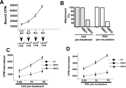
FIGURE 5.
HBD2 inhibits cellular entry of RSV. A, kinetics of 35S-methionine-labeled RSV (35S-RSV) attachment to A549 cells was examined by adding different amounts of virus (0.25 to 2 m.o.i. or 1.5 × 10⁵ to 12 × 105 cpm) to chilled A549 cells. Following attachment at 4 °C for 2 h, the cells were washed extensively, and the cell-associated radioactivity (in counts/min) representing the attached virus was measured by counting the cell lysate with a liquid scintillation counter. Each value represents the mean ± S.D. for three determinations. B, attachment of 35S-RSV (1 m.o.i., or 6 × 10⁵ cpm) to chilled A549 cells pretreated with HBDs (HBD1 or HBD2) or the virus particle preincubated with the HBDs was determined following attachment of the virus at 4 °C for 2 h. Following adsorption, the cells were washed extensively, and the cell-associated radioactivity (in counts/min) representing the attached virus was measured by counting the cell lysate with a liquid scintillation counter. The percentage of attachment was calculated as a ratio of the amount of radioactivity present in cells incubated with 35S-RSV in the presence of HBDs to the amount of radioactivity present in cells incubated with 35S-RSV alone. C, internalization of 35S-RSV (1 m.o.i., or 6 × 10⁵ cpm) into untreated (UT) or A549 cells pretreated with HBDs was determined following incubation of attached (2 h, 4 °C) virus at 37 °C for 0.5, 1, and 2 h. The cell-associated radioactivities (in counts/min) representing the internalized virus at different time points were measured by counting the cell pellet with a liquid scintillation counter. Each value represents the mean ± S.D. for three determinations. D, internalization of 35S-RSV (1 m.o.i., or 6 × 10⁵ cpm) into A549 cells following preincubation of the virus particle with HBDs was determined following incubation of attached (2 h, 4 °C) virus at 37 °C for 0.5, 1, and 2 h. The cell-associated radioactivities (in counts/min) representing the internalized virus at different time points were measured by counting the cell pellet with a liquid scintillation counter. Each value represents the mean ± S.D. for three determinations.

