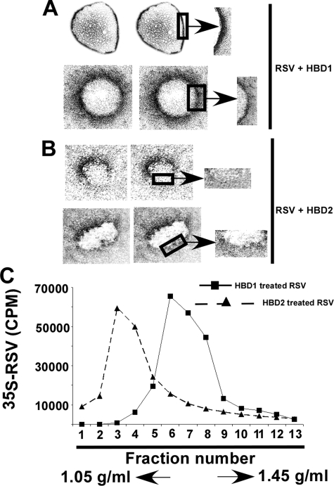
FIGURE 6.
HBD2-mediated disintegration of RSV envelope. Transmission electron micrograph of RSV virion particles following incubation with either HBD1 (A) or HBD2 (B) for 8 h. The boxed area representing a section of the viral envelope was magnified (denoted with arrows) to show the disruption of RSV envelope in the presence of HBD2. C, density gradient centrifugation of HBD1-treated (black squares and solid line) and HBD2 (black triangles and dashed line)-treated purified 35S-RSV virions on CsCl gradient (density gradient of 1.05–1.45 g/ml). Radioactivity (counts/min) present in the fractions was measured by a scintillation counter. Fraction 1 represents the lowest density and was recovered from the top of the gradient.

