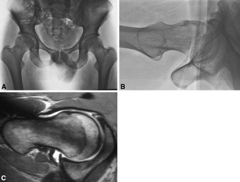
Fig. 3A–C.
(A) FAI is shown in a 34-year-old man with an apparently normal AP radiograph. (B) The nonspherical femoral head leading to reduced offset at the neck and predisposition to cam FAI is visible on the lateral radiograph. (C) The MRI scan confirmed the labral tear and chondral injury resulting from FAI. Reproduced with permission from Ganz R, Parvizi J, Beck M, Leunig M, Nötzli H, Siebenrock KA. Femoroacetabular impingement. Clin Orthop Relat Res. 2003;417:115.

