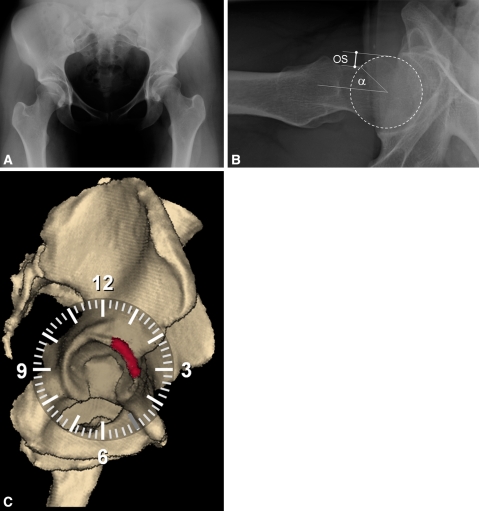
Fig. 2A−C.
This 26-year-old women presented with symptomatic anterior pincer FAI on the right side. (A) The anteroposterior pelvic radiograph reveals a bilateral coxa profunda without acetabular retroversion. (B) The femoral head demonstrated no signs of asphericity, the offset (OS) was more than 7 mm, and the alpha angle was 37°. (C) This snapshot shows the distribution of the sum of impingement zones for this patient for every possible combination of flexion, internal rotation, and adduction within a predefined maximum range (see text). The zones are located in the anterosuperior quadrant of the acetabulum when evaluating anterior FAI (red area).

