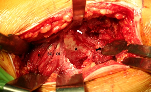
Fig. 2.
The acetabulum (A) is well-exposed after the complete capsulectomy. The tip of a Hohmann retractor is placed on the anterior wall of the acetabulum (white arrow); the proximal femur is retracted anteriorly (black arrow). The obturator internus (OI), superior gemelli (SG), and piriformis (P) are well-preserved.

