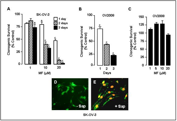
Fig. 3.

Effect of mifepristone on ovarian cancer cell clonogenic survival. In A, 250 SK-OV-3 cells were seeded in 6-well plates and exposed to 0, 1, 10 or 20 μM mifepristone (MF) for 1, 2, or 3 days. Following treatment the cells were cultured in regular growth medium for 7 days. At the end of the experiment the cells were fixed with 100% methanol and stained with 0.1% crystal violet. Colonies containing ≥ 50 cells were counted. The results are shown as number of colonies counted for the different experimental groups expressed as percentage of the control, which was considered to be 100%; a, P < 0.01 and c, P < 0.001 compared with control. In B, an experiment similar to that in A was done with OV2008 cells. The cells were exposed to 20 μM MF for 1, 2, or 3 days (c, P < 0.001 vs. control). In C, OV2008 cells were incubated with the indicated concentration of MF for 3 days, after which the cells were trypsinized, plated in a clonogenic survival assay, and their growth compared tocells that had not been treated with MF and whose growth was considered to be 100%. D and E show the results of a viability/cytotoxicity assay of SK-OV-3 cells upon exposure to MF. Cells were treated with 20 μM MF for 72 h and exposed to calcein AM plus EthD-1 in the absence (-Sap) (D) or presence (+ Sap) (E) of 0.1% saponin. Magnification × 200.
