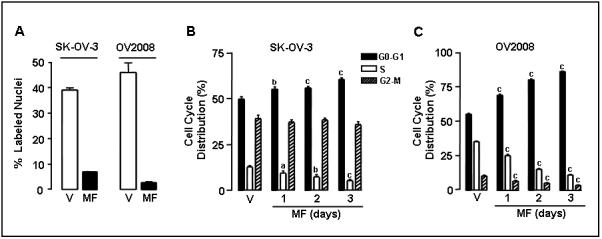
Fig. 4.

Effect of mifepristone on BrdU incorporation into DNA and on cell cycle kinetics in ovarian cancer cells. A, SK-OV-3 and OV2008 cells were cultured on multi-well slides and treated with either vehicle (V) or 20 μM mifepristone (MF) for 24 h. Cells were exposed to 10 μM BrdU for 12 h. At the end of the experiment, the cells were fixed with 4% paraformaldehyde, and BrdU incorporated into the DNA was visualized by immunocytochemistry using an anti-BrdU monoclonal antibody. BrdU positive nuclei were quantified using a microscope with a 100 × magnification and at least 500 cells were counted for each sample. The experiment was done in triplicate and the results are expressed as percent of BrdU positive cells. B, Logarithmically growing SK-OV-3 and OV2008 ovarian cancer cells were exposed to vehicle (V) or 20 μM MF for 1, 2, or 3 days. The percentage of cells in the G0/G1-, S-, and G2/M-phases of the cell cycle was determined by cytometric analysis of propidium iodide-stained cells. Values represent the means of two separate experiments performed in triplicate ± SEM. a, P < 0.05; b, P < 0.01; and c, P < 0.001 relative to the control group.
