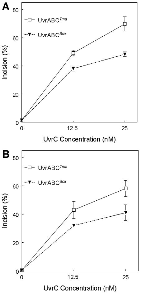
Figure 1. Incision of [3H]BPDE treated plasmids by UvrABCTma and UvrABCBca A. [3H]BPDE treated pTHQ008. B. [3H]BPDE treated pTHQB04.

Plasmids pTHQ008 and pTHQB04 treated with (+)-[3H]BPDE-anti (2 lesions/plasmid) were used as substrates for UvrA, UvrB and UvrC in a plasmid relaxation assay. Twenty fmol of DNA substrate were incubated with UvrABC in 20 μl of UvrABC buffer. Bca UvrA (2.5 nM) and Bca UvrB (62.5 nM) were held constant. Tma UvrC and Bca UvrC concentrations were varied as indicated. Form I and Form II plasmids were resolved by agaraose gel electrophoresis, quantitated and the average incision was calculated by applying the Poisson distribution and expressed as % of lesions incised. Means ± SD from quadruplicates reactions are plotted.
