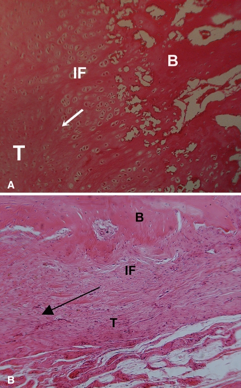
Fig. 5A–B.
A histologic section shows fibrovascular tissue in the interface (IF) between tendon and bone at 6 weeks, with a more robust fibrocartilage interface zone between the bone (B) and tendon (T) in the (A) growth-factor treated animals compared to (B) controls that received the collagen sponge alone. The arrow indicates the direction of pull of the tendon.

