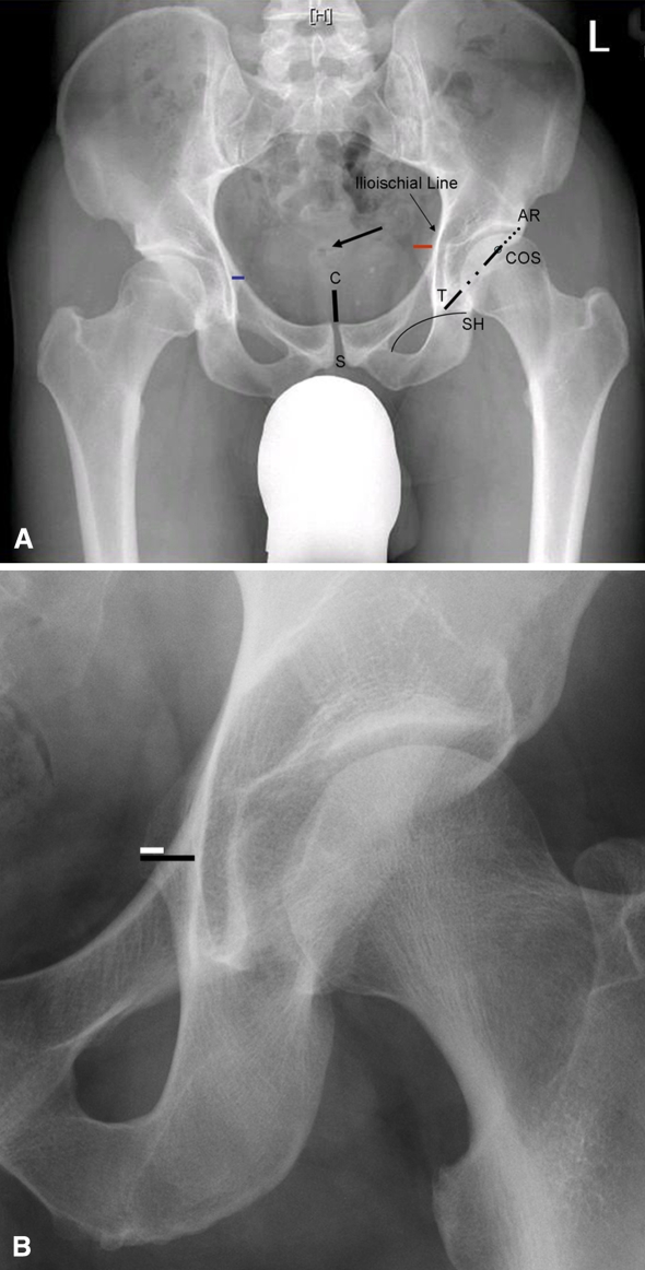
Fig. 2A–B.
(A) A pelvic radiograph shows the measurements obtained: symphysis (S) to coccyx (C) distance (black continuous line); sacrococcygeal joint (black arrow); COS, where the anterior and posterior walls cross (open circle); length of acetabulum (dotted black line and noncontinuous black line) from the lateral acetabular roof (AR) to the point that intersects the teardrop (T) with Shenton’s line (SH); PRIS 1 (blue line), the amount of ischial spine that was seen medially extending into the pelvic inlet (left side); and PRIS 2 (right side; red line), the distance from the ilioischial line to the tip of the ischial spine, even if its radiopaque shadow was situated behind the pelvic brim as shown. (B) A closer view of PRIS 1 (white line) and PRIS 2 (black line) is shown.

