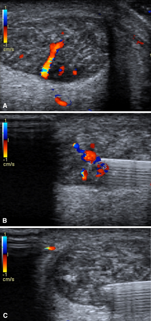
Fig. 3A–C.
Injection of a sclerosing agent is shown using Doppler ultrasound for guidance. (A) The presence of neovessels is detected in the Achilles tendon using color Doppler ultrasound before injection. (B) A 23-gauge needle is passed into the area of neovascularization under ultrasound guidance and the sclerosant is injected. (C) Ablation of blood flow within the neovessels is demonstrated after injection of the sclerosing agent.

