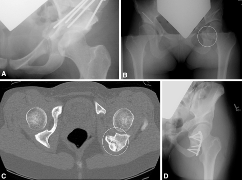
Fig. 2A–D.
(A) A radiograph obtained immediately after PAO shows an intact ischium. (B) An anteroposterior radiograph of the pelvis reveals an ischial fracture after periacetabular osteotomy. (C) The patient had a painful nonunion as confirmed on the axial computed tomography scan. (D) The postoperative anteroposterior radiograph shows the state after open reduction with internal fixation of an ischial fracture.

