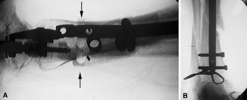
Fig. 2A–B.
(A) Intraoperative C-arm imaging of the distal tibia was performed to select one central hole as the pilot hole (arrows). Through the central hole (dotted circles) that overlapped the nail’s shadow, drilling of the medial cortex was performed only once. In this case, the upper central hole overlaps the shadow of the nail after its deformation. The pilot holes need not be perfect circles because they just served as guides in the anteroposterior plane to choose the closest one to the nail. (B) A final C-arm image shows distal locking of the tibia through the two locking holes.

