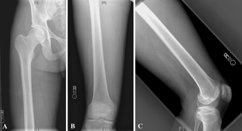
Fig. 2A–C.
(A) An AP radiograph of the right proximal femur taken 6 weeks after injury shows less extensive bone formation along the lateral aspect of the thigh. (B) Anteroposterior view of the right distal femur shows mild bone formation and a lateral (C) view of the right distal femur shows no evidence of bone formation along the anterior or posterior aspects of the thigh.

