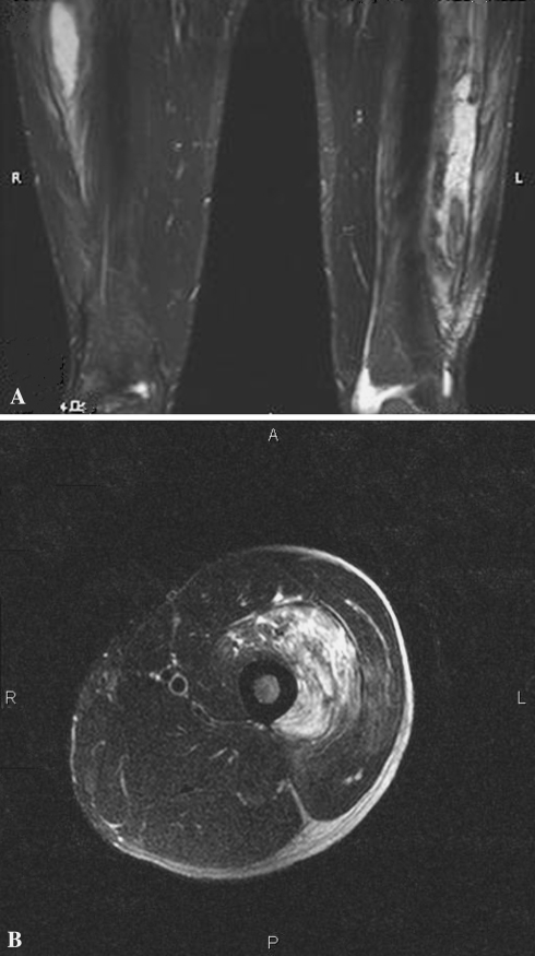
Fig. 3A–B.
(A) A T2-weighted MR scan shows the extent of myositis ossificans measuring approximately 13.3 × 2.3 × 3.5 cm in the left vastus lateralis muscle. There is a similar-appearing lesion in the upper right vastus lateralis measuring approximately 12.2 × 2.2 cm. (B) A T2-weighted axial MR scan shows a well-circumscribed multiloculated signal intensity focus in the vastus intermedius.

