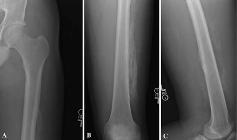
Fig. 4A–C.
(A) An AP radiograph of the left proximal femur taken 4 months after injury shows mature bone formation along the lateral aspect of the thigh. (B) Anteroposterior and lateral (C) views of the left distal femur also reveal mature bone formation both anteriorly and laterally 4 months after injury.

