Figure 8.
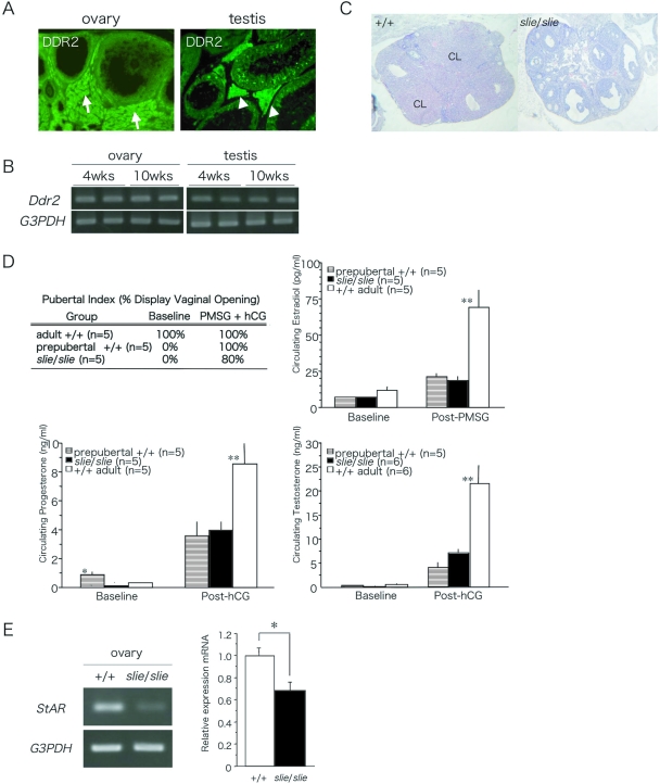
Localization and Expression of DDR2 in Adult Gonad and Responsiveness to Exogenous Gonadotropin Administration %A, DDR2 immunoreactivity was localized throughout the somatic cells of the ovaries and testes. Arrows indicate interstitial cells between follicles (left panel), and arrowheads indicate Sertoli cells (right panel). B, Ddr2 was expressed in prepubertal and adult ovary and testis. C, Ovarian sections stained with hematoxylin and eosin after exogenous gonadotropin stimulation to 10-wk-old wild-type and slie mutant mice. D, Presence of vaginal orifice opening and serum estradiol levels 24 h after PMSG and serum progesterone levels 24 h after PMSG + hCG in female mice, and serum testosterone levels 24 h after 10 hCG in male mice (*, P < 0.05; **, P < 0.01). CL, Corpus lutea. E, Semiquantitative RT-PCR of StAR after gonadotropin administration in wild-type and slie mutants.

